
Further Reading - Gamma Rays
Gamma Knife
Decay of Cobalt-60
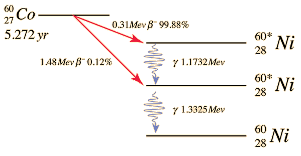
The gamma rays are produced by the beta decay of cobalt 60 to nickel 60. Cobalt decays to Nickel, an electron and an anti electron neutrino. The decay produces two high energy photons, 1.17 and 1.33 MeV.
Cobalt-60 has a half life of 5.272 years, which means it takes that long for the source to reduce in activity by half.[G14]
Image 1: Decay of Cobalt-60. Source (Hyperphysics)
How gamma knife treatment is targetted

Image 2: Isodose curves for a coronal slice of a brain. Source (Optimisation of gamma knife radiosurgery [G16])
A 3D coordinate system is used to locate targets in stereotactic space (Z = axial, X = Sagittal and Y = coronal). The skull is then measured with the frame and the measurements are used by gamma software to formulate the treatment and how the beams should be angled. The dose is calculated in a 31 x 31 x 31 matrix of points centred on the target. [G15]
This is combined with the results of a brain scan to determine where exactly the treatment needs to be.
Isodose curves can be used to optimise the treatment plan. A 50% isodose curve is a curve that contains all of the pixels that receive 50% or more of the dose delivered. This can be seen in image X, where the 50% (yellow) and 30% (green) curves can be seen, this image is a coronal (y-axis) slice of a brain. [G16]
How gamma knife dosage is calculated
The dose calculation for one beam depends on the skull configuration, transverse radiation (for a specific collimator size) and the dose reference in a water phantom. This is then calculated for all sources, which are summed for each point in the 31x31x31 matrix. The dose is the sum of the contributions of each ‘shot’ of radiation delivered at the possible shot sizes (4mm, 8mm, 14mm and 18mm). The equation for this is then:

Where W is the possible shot sizes, S is the set of shots considered, t_s is the length of time for delivery,
is the dose delivered to the pixel (i,j,k) by the shot of width w centered at .
is found by simulating a shot centered at the middle and finding the dose delivered in the x,y and z directions at a given distance, then taking the average of these.[G16]
Gamma Cameras
Video 1: How a gamma camera works. Source (YouTube, video by Physics@LSC)
The gamma produced is then detected by the gamma camera. A gamma camera works in the following way[G17]:
The camera is made up of collimators, scintillators and photomultiplier tubes. The collimator is just a configuration of lead, which block out gamma rays if they are at an angle. This means that the origin of the gamma can be determined because it is exactly below where it is detected.
The scintillator is a material that absorbs the gamma rays and emits visible light. This is useful for the photomultiplier tube which detects visible light. The light kicks out an electron in the photomultiplier tube, which then results in an electrical signal. This can then be picked up by a computer to make an image.
The video on the left is an animation of how this occurs.
Medical Tracers
Decay of Technetium-99m

The decay of Technetium-99 is interesting because it decays from a metastable state to a stable state, giving off a photon with energy 140.5keV most of the time although the decay can have different results.
Image 2 shows details of this more clearly. The half-life of this decay is 6.03 hours, which is relatively long for the type of decay but it makes it a good candidate for medical imaging.
The half-life dictates how long it will take for the source to halve in activity, so a half-life that is relatively short is ideal for medical tracers. A half-life that is too long would increase the dosage which is not ideal but a half-life that is too short would not give a good image.[G18]
Image 3: Decay of Technetium-99m. Source (Hyperphysics)
PET Scans
Antimatter

Antimatter is essentially matter but with one key difference, the charge and magnetic moment are flipped.
Antimatter was discovered by mathematician Paul Dirac when he tried to combine quantum mechanics with relativity. Dirac attempted to relate the two using 2D numbers (matrices) and discovered his equation allowed for positive and negative energies, ie matter and antimatter.
The existence of which was discovered in 1923, when Dmitry Skobeltzyn tried to look for gamma rays and found some electrons moving opposite to their expected paths. [G19]
The image on the left shows matter with it's corresponding anti matter part. Antimatter is denoted by a line above the symbol.
Image 4: Matter and antimatter split into quarks and leptons. Source (Annenberg Learner, Image by Fermilab)
Antimatter Annihilation

Image 5: Electron and positron annihilation. Source (Wikimedia, Image by Jabberwok)
When an electron and a positron meet, they will annihilate each other and produce two gamma rays with energy 511keV.[G20] Said gamma rays are aligned in opposite directions, so momentum and energy is conserved.
Image 4 shows a Feynman diagram of this interaction, the straight lines are the leptons (electron and positron) and the wiggly line is a boson (photon) of gamma radiation. Theoretically this interaction can occur both ways.