Thermography
When we are ill, one of the body’s natural responses is to raise its body temperature. This change in temperature can therefore be a useful indicator of sickness. This is the basic principle of Infrared Thermography. Much like other medical imaging techniques, it forms an image either from the body’s emitted radiation (MRI & Microwave Tomography) or using radiation which has passed through the body.
What is Thermography?
A Thermogram which compares a normal hand to that of a hand suffering from Raynoud's Syndrome.
Image Source (Welcomeimages)
Thermography, like the former, is a painless, passive, non-invasive, non-contact imaging technique which can form an image using a heat map, much like those seen in the weather forecast, of the Infrared light (IR) given off by the surface of the human body. With these detected IR waves and statistical calculations on a computer it is possible to identify regions of the body which are abnormal in temperature which can be used an effective diagnostic tool in medicine.
How does it work?

Thermogram of a normal breast showing a symmetric distribution of heat. Image Source (ThermologyOnline)

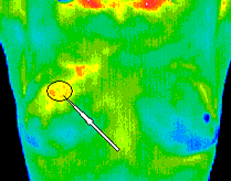
Thermogram showing the early stages of a malignant tumour in the breast.Image Source (ThermologyOnline)
Safety
Similar to Microwave Tomography, this imaging is passive and so relies solely on the energy radiated from the human body. This means that it is naturally safe to take the scan as it is just like getting a picture taken!
The camera is sensitive to temperature changes of 0.05°C and so, for example, detects energy changes of around 4J in the breast. This is a negligible number when compared to the tea scale and so we will say that it would not be able to heat any cups of tea.
Temperature fluctuations within the body come from different metabolic processes. These internal temperature fluctuations cause the temperature of the human skin to vary slightly across an area. Human skin has an average temperature of 27°C which can be shown to emit most of its IR light at a wavelength of 9.63μm by calculation. The hottest object in a room will be the brightest. Therefore, when the human body is fighting sickness or has another condition such as a tumour or disease, it will produce clear recognisable heat patterns distinct from its surroundings which can be detected using an Infrared Camera. This camera is tuned to view a range of wavelengths between 8-12μm. This is known as the “body infrared ray band” [IR4]
This camera usually contains a microchip made of Silicon wafer which has the capability of detecting IR waves to an accuracy high enough to distinguish a temperature fluctuation of 0.05°C on the surface of the body. This camera is able to take live readings of the IR waves coming from the body and when paired with a computer processor can form 2-D images of the body in view. These images are then given “false colour” to produce a heat map, where higher temperatures are depicted with lighter colours and lower temperatures with darker ones.
Many clinical illnesses have been detected using this method of imaging. One of the common identifiable patterns imaged is that of a patient suffering from Raynoud’s Syndrome whose fingers and toes take around 10 minutes to return to normal body temperature after they have been exposed to the cold.
Tumours in the breast can also be detected at early stages thanks to their characteristic heat pattern displayed on the Thermogram (images taken using the IR Thermography imaging technique). An unhealthy breast tissue will produce a higher temperature that its surroundings. Abnormal results from a breast thermogram are taken to be a significant risk marker for the existence of a breast tumour. [IR5]

Right: The energy change which can be detected inside the body by a Thermogram is so small that it won't be able to heat a cup of tea.
